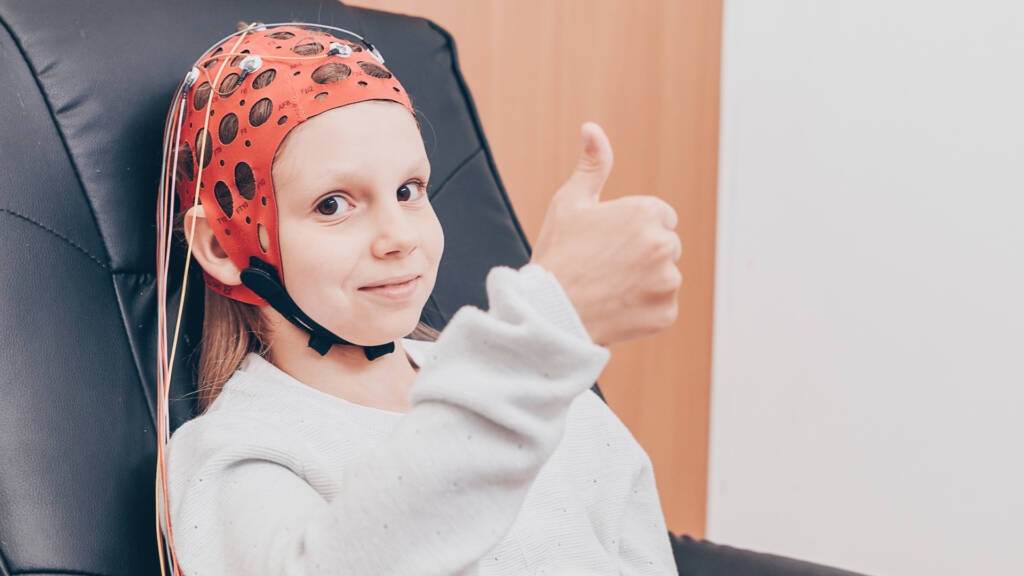
Brain Mapping helps us understand what is going on inside your brain. Brain Maps, also known as a Quantitative EEG or QEEG, record you brain’s electrical activity. Before starting Neurofeedback it is essential to first complete a Brain Map to guide the therapeutic approach. Based on your brain map, we will find areas you can improve such as focus, executive functioning and impulse control. We will not be attaching labels to you because for many people labels are hard to shed and become part of one’s identity.
What to expect during a Brain Map…
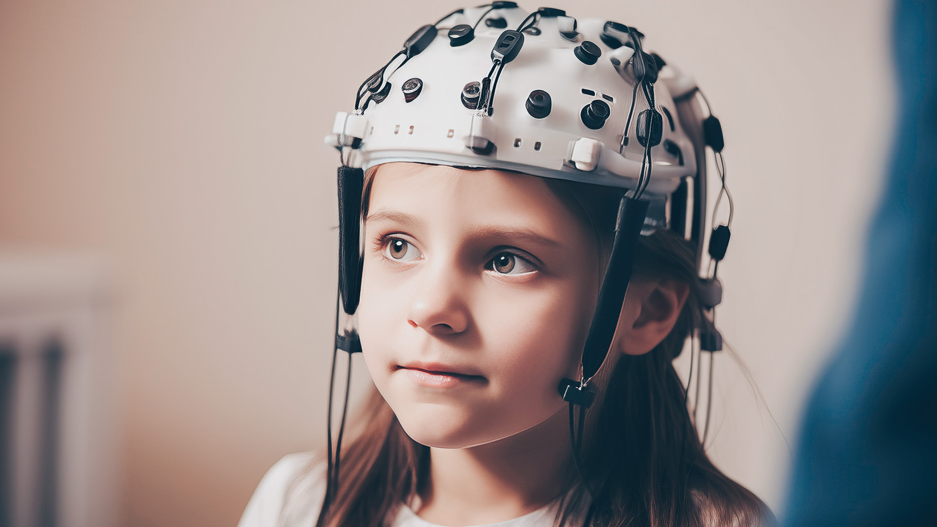
This procedure takes place in our office for a total of 20 minutes. An elastic net cap with 19 sensors will be placed on your head with gel to ensure brainwave activity is recorded. Once complete, this data will then be interpreted alongside your intake and a personalized plan will be created specifically for your goals based on the outcome from the Brain Map. We generally repeat Brain Maps around Neurofeedback session 20, so we can visually see the progress and dynamic change your brain has made.
Who Brain Mapping can help…
Children & Adolescents
We can help with…
Academic Performance
ADHD
Anger
Anxiety
Autism
Spectrum Disorder
Behavioral Issues
Concussion & Traumatic Brain Injury
Depression
Intellectual & Developmental Delay
Learning Disabilities
Obsessive Compulsive Disorder
PTSD
Sleep Issues
Stress Response
Trauma
Adults
We can help with…
Alzheimer’s
Dementia
Depression
Anxiety
Memory
PTSD
Concussion/ TBI
Stroke
Brain Fog
Learning Disability
Stress Response
Intellectual & Developmental Delay
Sleep Issues
How it works…
An elastic net cap with 19 sensors is placed on the head with some gel so that the brainwave activity can be measured. There is no piercing of the skin. Brainwaves are then recorded for 5-10 minutes with eyes closed and again with eyes open. This data is then compared to one or more normative databases. The results specify where there are problems with dysregulation and that leads to a personalized neurofeedback protocol. For example, if an ADHD individual is making too much sleepy theta waves in their left frontal lobe, that might lead to placing the sensor there during the neurofeedback sessions and training the brain to make less theta there. The Brain Map reveals the specific type of brain dysregulation which underpins the diagnosis.


